OPTICAL APPEAL
A novel non-invasive skin imaging tool is changing the way cancers are diagnosed at the National Skin Centre.
A lesion on his forehead that Mr Lim had for more than a year suddenly started bleeding. Alarmed, Mr Lim’s family members urged him to get it examined at the National Skin Centre (NSC).
It turned out to be the right thing to do, as Mr Lim was diagnosed with basal cell carcinoma, one of the most prevalent types of skin cancer, both in Singapore and globally.
Naturally, Mr Lim, a retiree in his 70s, was anxious before his diagnosis, but it helped that he did not have to wait long to get the results. In fact, Mr Lim received his results on the same day.
Until recently, diagnosing basal cell carcinoma was a time-consuming process that could take up to two weeks, including waiting time for a biopsy appointment and a histopathology report. Now, thanks to a faster and less invasive diagnostic imaging method introduced at the NSC, the diagnostic process has been reduced to same-day diagnosis in most cases.
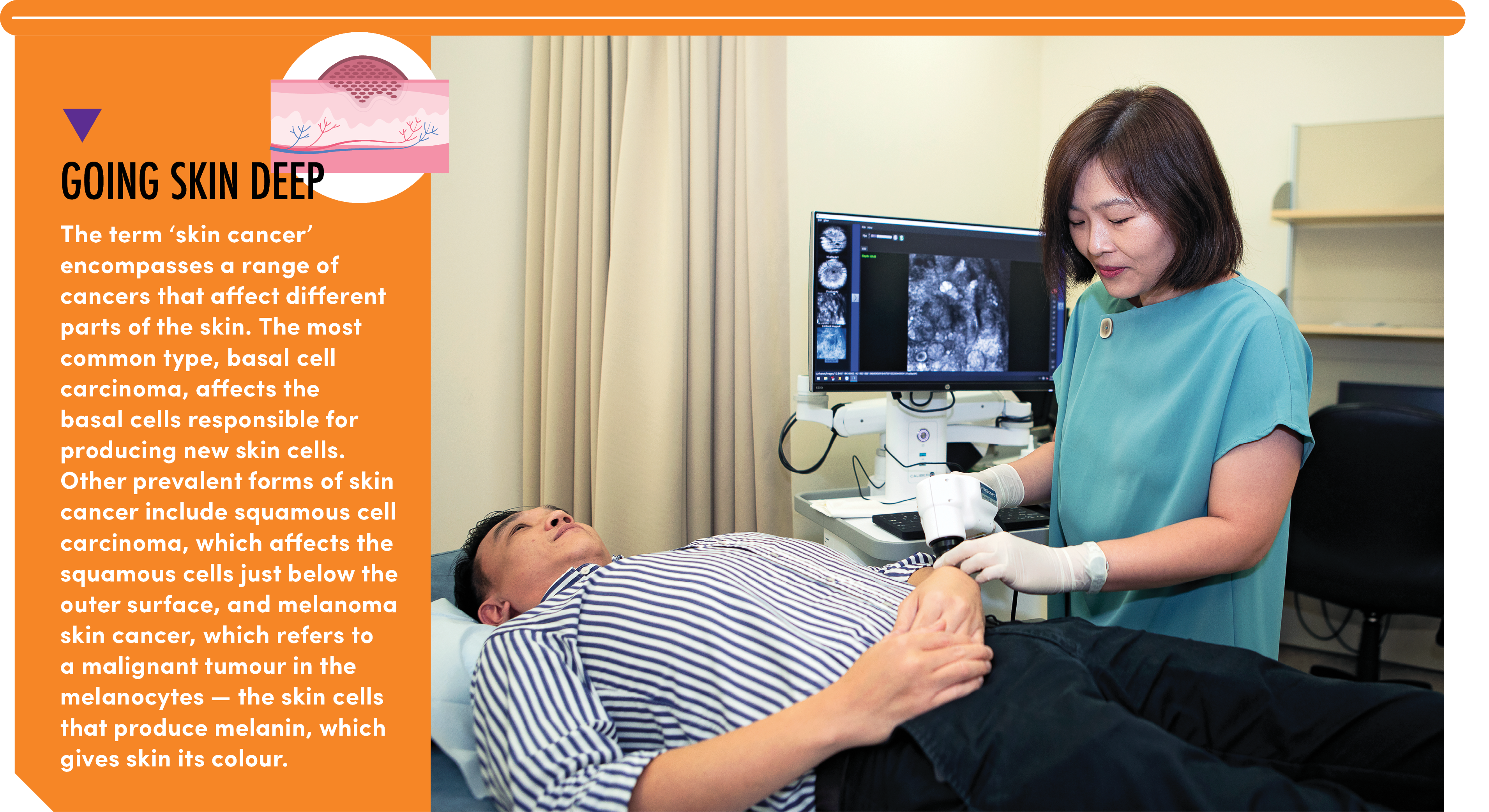
The method, known as Reflectance Confocal Microscopy (RCM), was introduced as a clinical service at the NSC in November 2021*. RCM is considered a ground-breaking skin cancer imaging and diagnostic technique in Singapore, where skin cancers rank as the sixth most common form of cancer. Its introduction at the NSC marks the first application of this method in Southeast Asia, following its successful implementation in Europe, the United States, and Australia.
ZOOMING IN ON RCM
RCM is non-invasive and painless. It employs a low-power laser to produce a black-and-white image of skin lesions. “The process was comfortable and not at all frightening,” recalls Mr Lim, who underwent the procedure in April this year.
“It felt almost like undergoing an ultrasound. The doctor directed the device towards the lesion, and within 15 to 20 minutes, we were done. All I had to do was just lie on the bed.”
An added advantage of RCM is that the procedure is safe for most patients, including pregnant women.
On the other hand, the traditional method for diagnosing skin cancers is through a biopsy. It is an invasive procedure in which a small sample of skin is extracted for further investigation. Removing the sample requires a dose of local anaesthetic, and stitches are needed to close the wound. “After a biopsy, there may be bleeding, bruising, and a risk of infection. All biopsies result in scarring,” notes Dr Chuah Sai Yee, Senior Consultant at the NSC and Consultant-in-Charge of
the Pigment and Skin Imaging Clinic.
After a dressing is applied, patients are given instructions on wound care. They would have to
return in about two weeks to have the stitches removed — and to receive their diagnosis.
Shortening this procedure is one of the primary advantages of RCM, explains Dr Chuah. By delivering a diagnosis within minutes, patients are spared the emotional distress of having to wait for days for their test results. The reduced diagnostic time with RCM also enables treatment, if needed, to be started promptly.
These factors appealed to Mr Lim, who was given a choice between a traditional biopsy and
RCM. “I chose RCM because of the faster diagnosis. We could get certainty whether the lesion was cancerous, or not, within a day, and if it was, we could arrange for it to be surgically removed.” Mr Lim had his lesion removed on the same day as his surgeon was available to perform the procedure.
"With RCM, we can assess all these lesions at once, eliminating the need for multiple biopsies"
- Dr Chuah Sai Yee, Senior Consultant, Consultant-in-Charge, Pigment and Skin Imaging Clinic, National Skin Centre
RCM is also more cost-effective as compared to a biopsy and histology report for a similar lesion. “RCM is particularly useful for patients with multiple lesions that need to be examined for cancer,” says Dr Chuah. “With RCM, we can assess all these lesions at once, eliminating the need for multiple biopsies.”
RCM can also be used to diagnose other skin conditions, including pigmentary disorders like vitiligo and melasma, as well as skin infections and inflammatory skin conditions.
Given the multiple advantages of RCM, why is it not used as the default method of diagnosing skin cancers? “Like all skin imaging tools, RCM has its limitations,” says Dr Chuah. “RCM may not be able to visualise deeper skin structures and may be less effective on lesions that are ulcerated, bleeding, or those with thick scales.”
However, a medical professional can determine the best approach of finding out whether a lesion is benign or cancerous. “When the results of a RCM are unclear, we will carry out a biopsy to be certain.”
Accuracy is crucial in medical settings, and according to Dr Chuah, RCM is a dependable iagnostic tool. Based on data analysis collected over three years*, RCM has a success rate of between 80 and 90 per cent in diagnosing skin cancers.
Dr Chuah Sai Yee (centre) and research staff with the RCM at NSC's Skin Imaging Clinic.
That is why RCM is part of a wider suite of diagnostic tools, which include biopsies and dermoscopy examinations. The latter is performed with a dermatoscope, a handheld visual aid used to examine skin lesions in greater detail. Regardless of the tool used, Dr Chuah advises those with unusual skin lesions to seek medical advice as soon as possible — the earlier, the better.
Mr Lim echoes this advice, “If you notice anything suspicious about your skin, have it examined by a healthcare professional. I am glad I had my lesion checked and promptly removed. It left me with peace of mind.” LW
Read the e-Book of the magazine
hereRead the PDF of the magazine here